Bruker SENTERRA II

Bruker SENTERRA II – Confocal Raman Microscope
Bruker SENTERRA II is a compact and fully automated confocal Raman microscope designed for fast and precise material identification, analysis, and imaging. It combines research-grade performance with ease of use, enabling Raman imaging, 3D analysis, and reliable material identification. With SureCal™ technology, the system continuously maintains high wavenumber accuracy without the need for frequent manual calibration.
ADVANTAGES
APPLICATIONS
TECHNICAL SPECIFICATIONS
VIDEO
Advantages
- Fully automated Raman microscope
- Up to 4 integrated lasers with one-click switching
- SureCal™ continuous calibration technology
- High wavenumber accuracy
- Fast 2D and 3D Raman imaging
- Automated workflows for simplified operation
- Automatic self-diagnostics and OQ/PQ testing
- Low maintenance requirements
- Class 1 laser enclosure with safety interlock
- Advanced material identification and analysis tools
- Support for GMP and 21 CFR Part 11 requirements
Applications
SENTERRA II is suitable for a wide range of industrial, research, and analytical applications:
- Pharmaceuticals – analysis and mapping of active ingredients in tablets
- Materials science – batteries, graphene, silicon, and other advanced materials
- Polymers and plastics – material identification, filler analysis, and component distribution
- Microplastic and particle analysis
- Quality control and failure analysis
- Forensic analysis and identification of unknown substances
- Art conservation and restoration
- Biological and life science research
- Coating, interface, and multilayer analysis
- Research and development
Technical Specifications
- Technology: Confocal Raman microscopy
- Laser excitation: Up to 4 integrated lasers, depending on configuration
- Calibration: SureCal™ continuous automatic calibration
- Raman imaging: 2D and 3D Raman imaging
- Microscopy: Confocal µ-Raman analysis
- Automation: Fully automated hardware and optical component adjustment
- Validation: Automated OQ/PQ tests
- Software: OPUS
- Data analysis: Material identification, Cluster ID, Find Particles, and Adaptive K-means
- Laser safety: Class 1 enclosure with safety interlock
- Options: FT-Raman module, Raman probes, various sample holders, liquid cells, and temperature-controlled stages
- Life science configuration: Optional inverted microscope configuration for cell and tissue analysis
Product Related
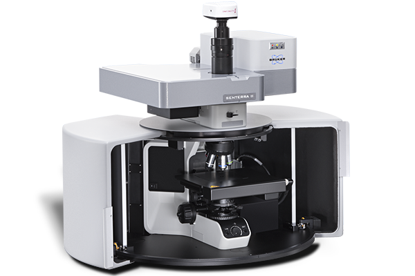
Bruker SENTERRA II
Bruker SENTERRA II – Confocal Raman Microscope Bruker SENTERRA II is a compact and fully automated confocal Raman microscope designed…
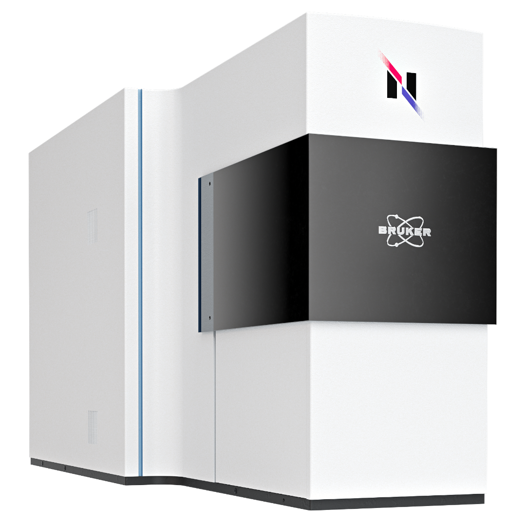
Bruker Nanophoton Series Microscopes
Nanophoton Raman Microscopes Nanophoton Raman microscopes combine high-resolution Raman spectroscopy with advanced galvanometer scanning technology for fast and precise chemical…